Runwen Yao
Biomolecular condensates · mesoscale organization · cellular function
Home
Research
Research Overview Theme I: Mesoscale regulation Theme II: Physical principles Theme III: Quantitative tools
PublicationsPubs CV Gallery
← Back to Gallery

Scientific Imaging

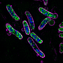
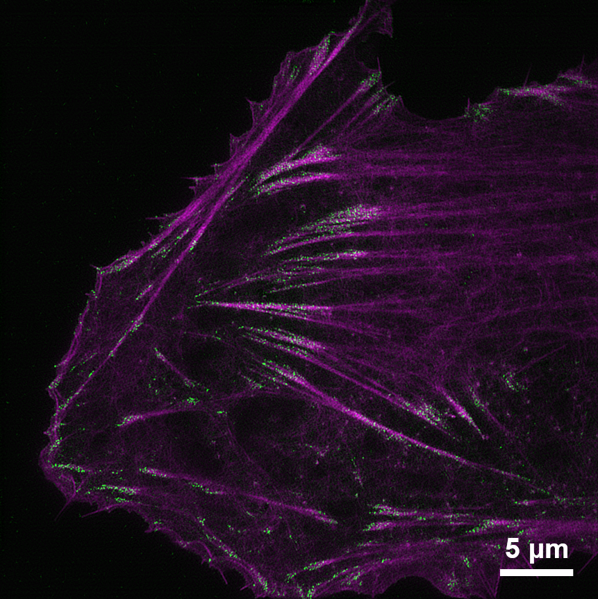
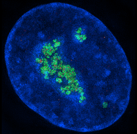
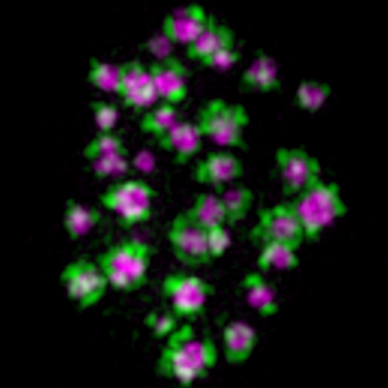
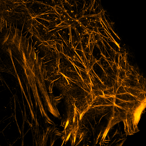
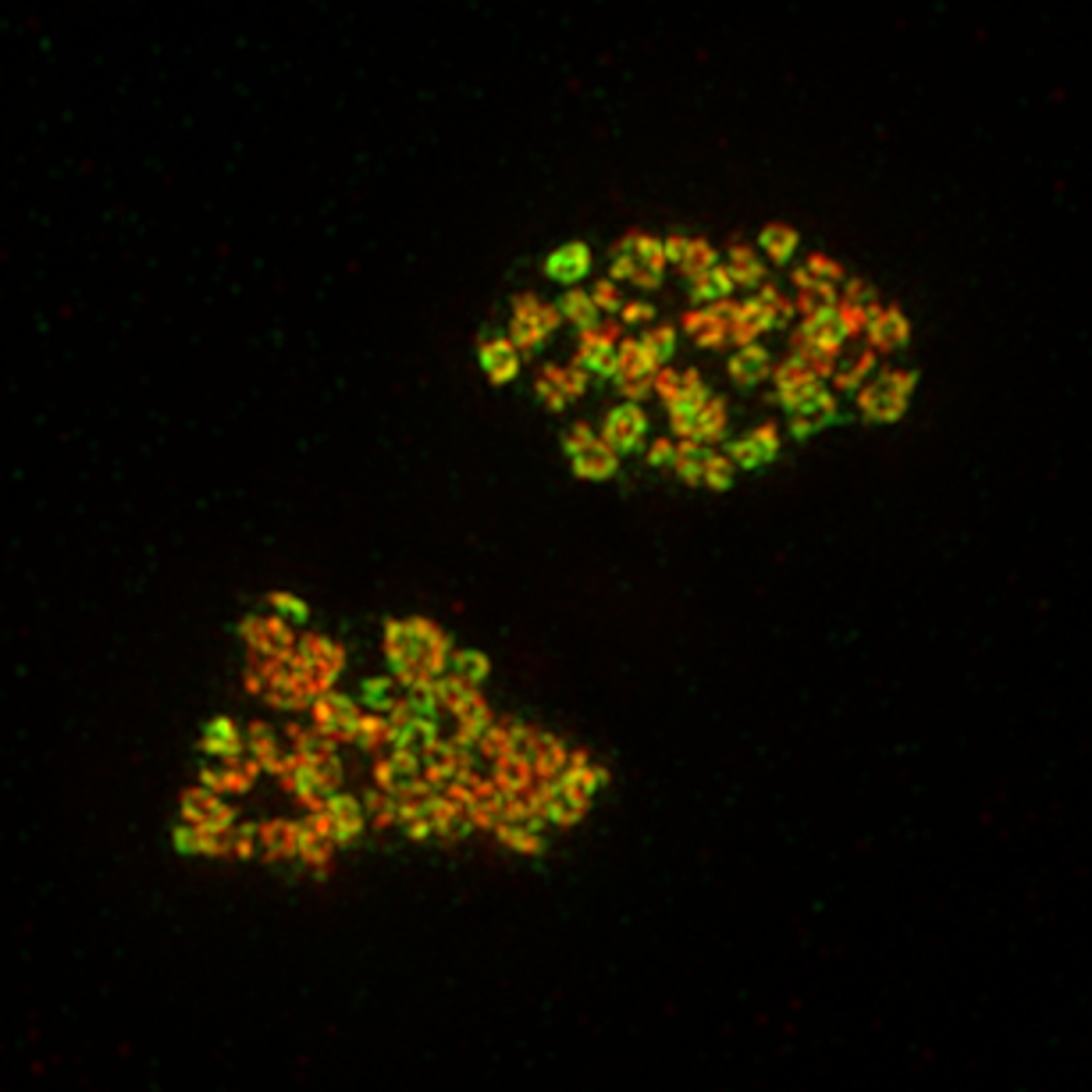
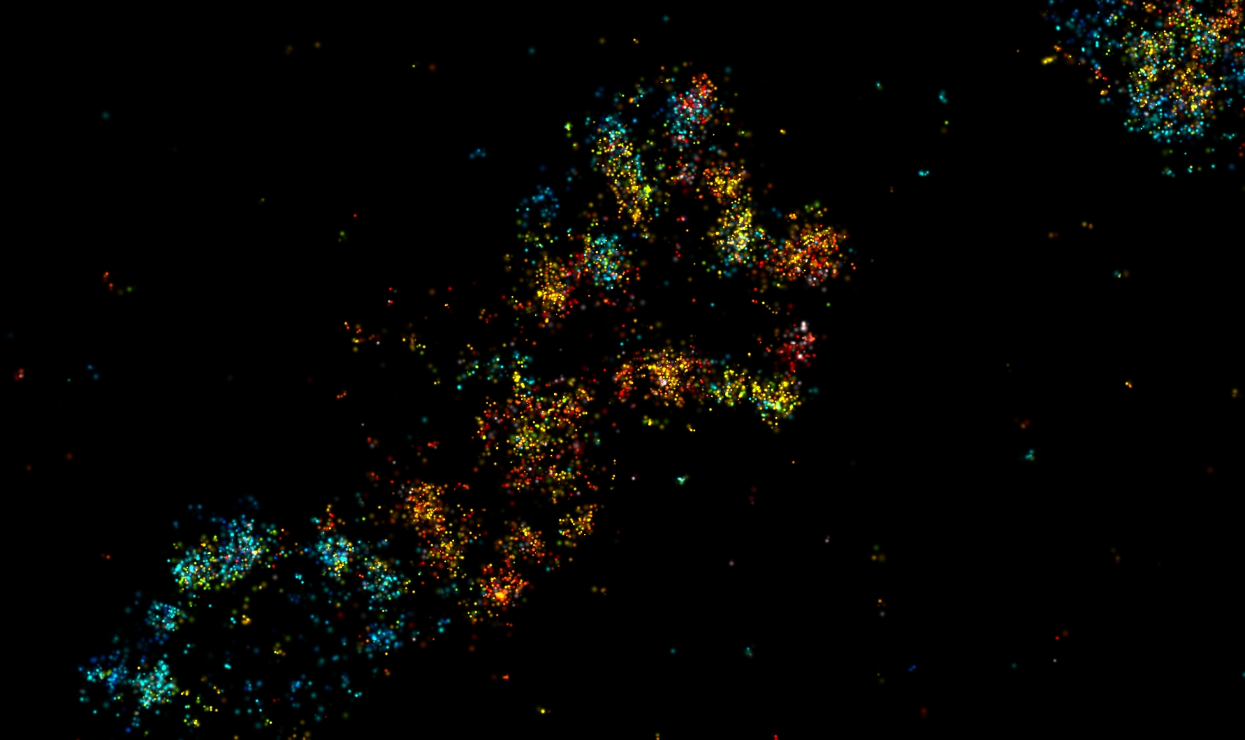
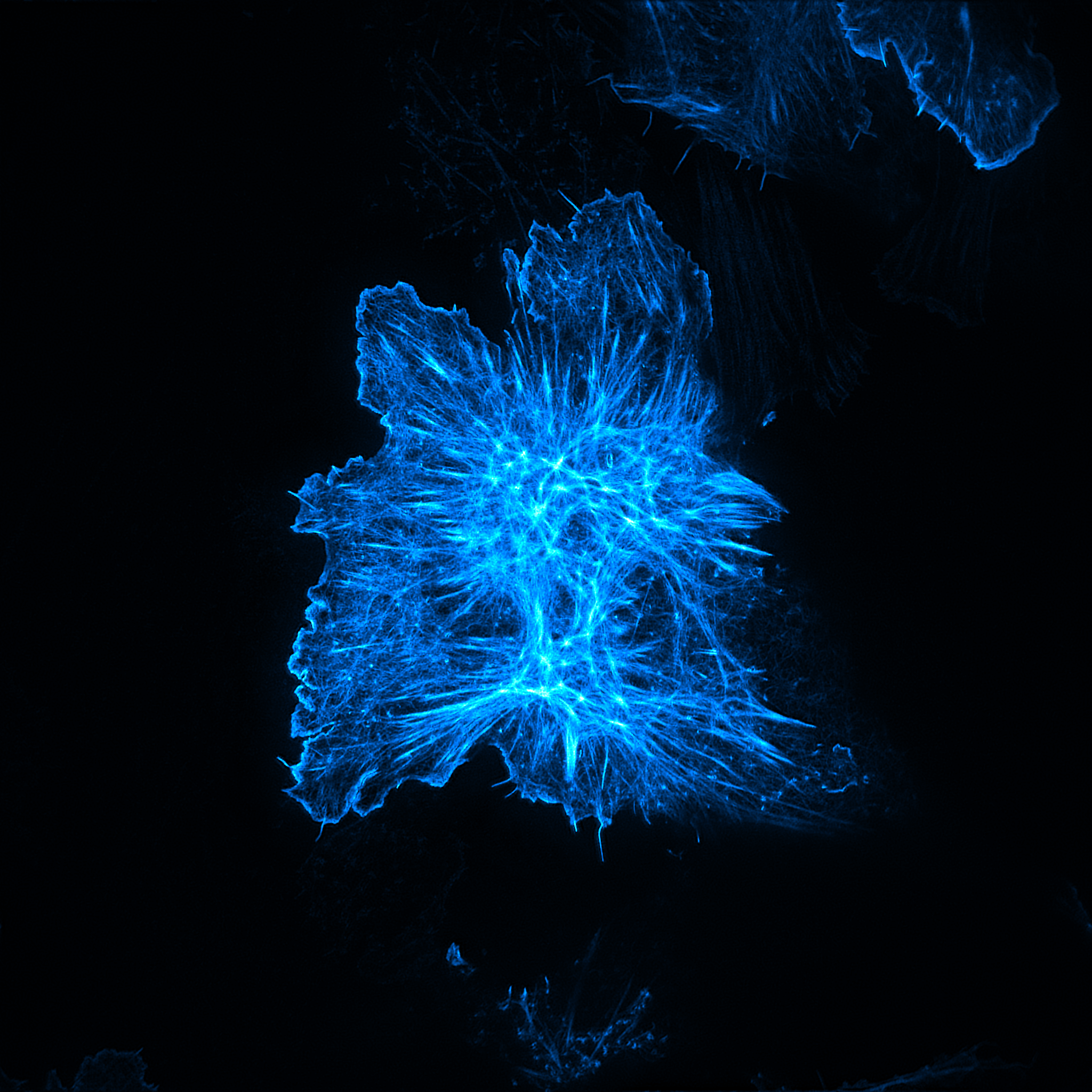
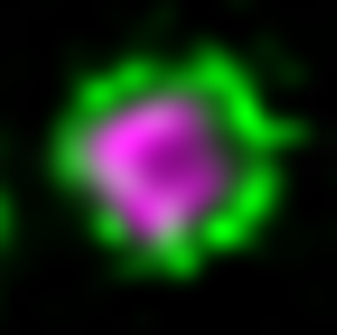
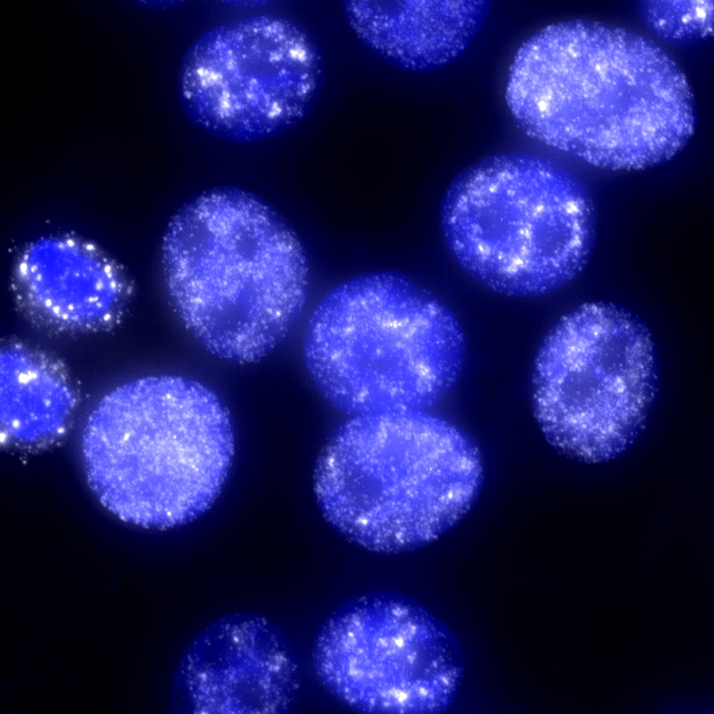
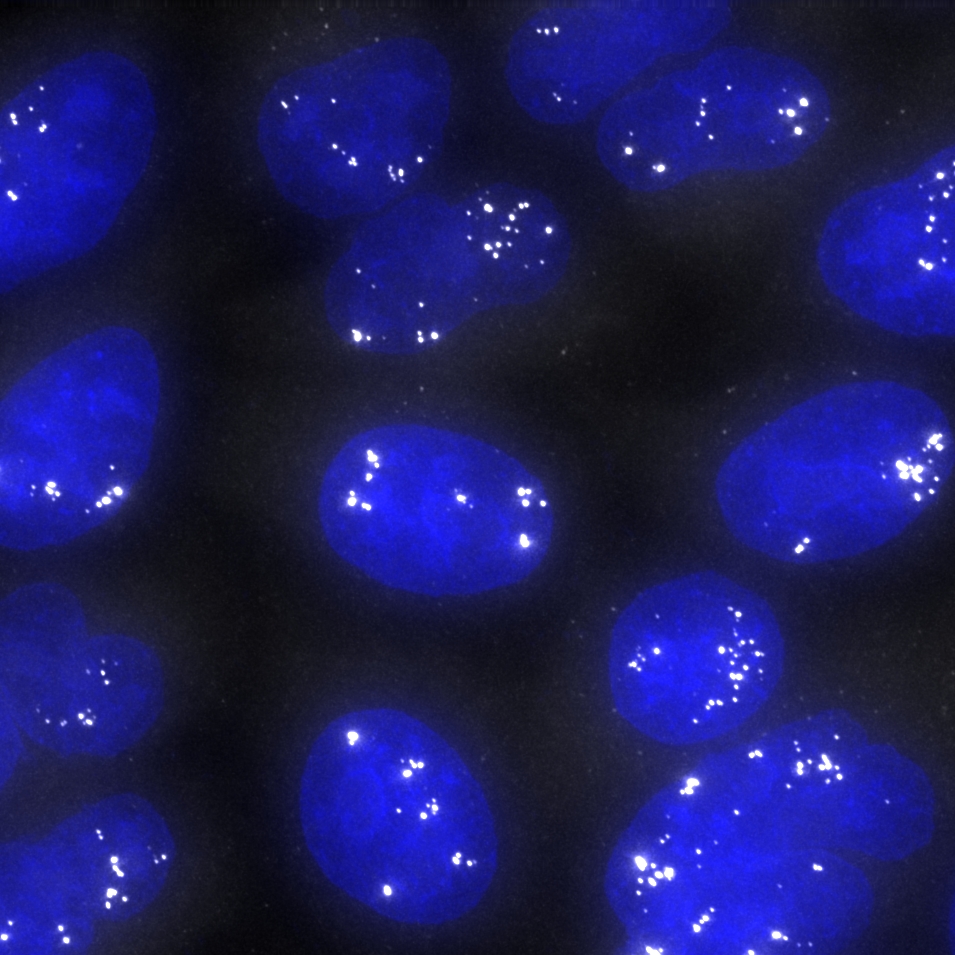
Microscopy, condensates, and cellular organization viewed through the experimental systems that shape my research.

Figure thinking placeholder gallery image
3D-SIM

Vibrio parahaemolyticus

PKMO FX (magenta), protein of interest (green), and DAPI (blue)

light-sheet microscopy

72 hpf zebrafish

RNA (magenta), Nucleoli (green), Pol II (red), Nuclei (blue)

Figure thinking placeholder gallery image
TIRF-SIM

Actin and Nck

LifeAct-labeled actin (magenta), Nck (green)

Internal organization placeholder gallery image
3D-SIM

Nucleolar organization

FBL (green), RPA194 (red), Nuclei (blue)

Internal organization placeholder gallery image
3D-SIM

FC/DFC in the nucleolus

FBL (green), RPA194 (magenta)

Wide placeholder gallery image
TIRF-SIM

Actin cytoskeleton architecture

Phalloidin-labeled actin (blue)

Soft placeholder gallery image
STED

Paraspeckle spatial organization

NEAT1_5′/3′ (green), NEAT1_mid (magenta)

Figure thinking placeholder gallery image
smFISH

MALAT1

MALAT1 (white), DAPI (blue)

Internal organization placeholder gallery image
smFISH

NEAT1

NEAT1 (white), DAPI (blue)

Internal organization placeholder gallery image
TIRF-SIM

Actin cytoskeleton in U2OS

LifeAct-labeled actin (yellow)

Figure thinking placeholder gallery image
3D-SIM

DDX21 ring structures

DDX21 (green), FLAG-tagged protein (red)

Notebook placeholder gallery image
SMLM

Nascent pre-rRNA

Color encodes depth

Runwen Yao HHMI / UT Southwestern Medical Center
© Runwen Yao. Please contact me before reusing images or figures.